Home

cleric Drepturi de autor gudron protein electron microscope aplecare Sud etichetă

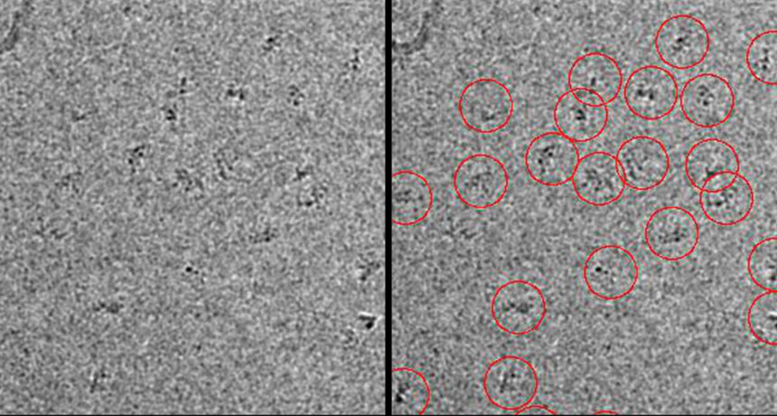
Making the nanobody big again for Cryo-EM

Imaging proteins at the single-molecule level | PNAS

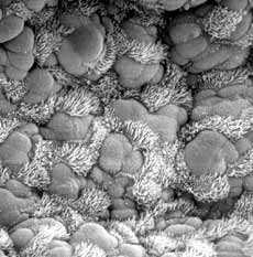
Scanning electron microscope observations of protein bodies in the... |  Download Scientific Diagram
Scanning electron microscope observations of protein bodies in the... | Download Scientific Diagram

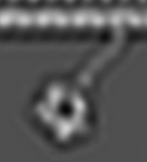
First images of motor proteins in action - 2015 - Wiley Analytical Science

Supercool Protein Imaging Gets the Nobel Prize | Quanta Magazine

Chilled proteins and 3-D images: The cryo-electron... - Nuadox |  Innovation, science and tech news
Chilled proteins and 3-D images: The cryo-electron... - Nuadox | Innovation, science and tech news

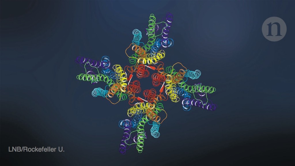
Cryo-electron microscopy shapes up

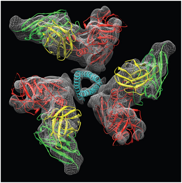
HIV Protein Strikes a Fleeting Pose | National Institutes of Health (NIH)

Cryo-electron microscopy reaches resolution milestone

Explainer: What is cryo-electron microscopy | News | Chemistry World

What is Electron Microscopy? - UMASS Medical School

Imaging proteins at the single-molecule level | PNAS

Biologists seek help to 'see' itty-bitty molecules in 3-D

Transmission Electron Microscopy as an Orthogonal Method to Characterize  Protein Aggregates - Journal of Pharmaceutical Sciences
Transmission Electron Microscopy as an Orthogonal Method to Characterize Protein Aggregates - Journal of Pharmaceutical Sciences
Determining Protein Structure with Electron Microscopy

Cryo-electron microscopy reveals how protein crystals grow

Electron microscope image, prion protein fibrils stock photo

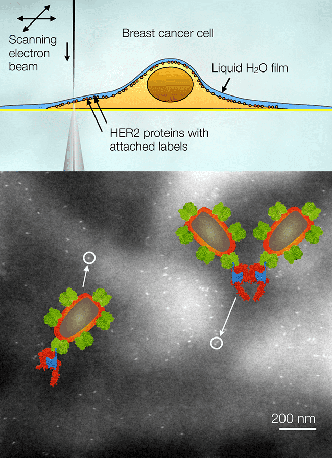
Invited Lecture ELECTRON MICROSCOPY OF MEMBRANE PROTEINS IN EUKARYOTIC  CELLS IN LIQUID - ISM2016 (Microscopy)
Invited Lecture ELECTRON MICROSCOPY OF MEMBRANE PROTEINS IN EUKARYOTIC CELLS IN LIQUID - ISM2016 (Microscopy)

Near-atomic resolution of protein structure by electron microscopy holds  promise for drug discovery | National Institutes of Health (NIH)
Near-atomic resolution of protein structure by electron microscopy holds promise for drug discovery | National Institutes of Health (NIH)
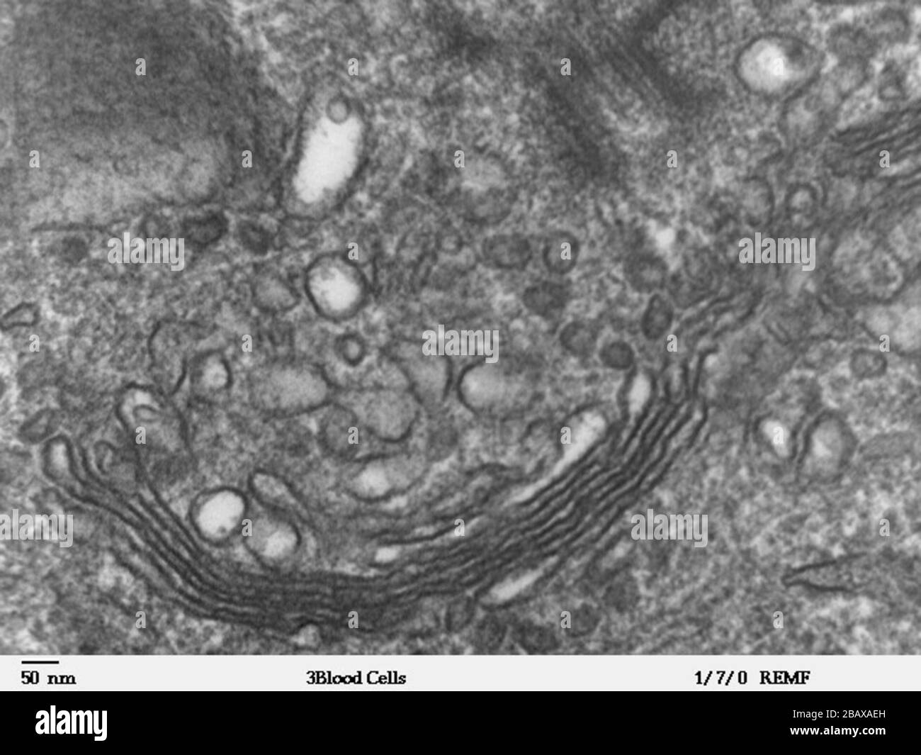
High magnification transmission electron microscope image of a human  leukocyte, showing golgi, which is a structure involved in protein  transport in the cytoplasm of the cell. JEOL 100CX TEM;  http://remf.dartmouth.edu/imagesindex.html http://remf ...
High magnification transmission electron microscope image of a human leukocyte, showing golgi, which is a structure involved in protein transport in the cytoplasm of the cell. JEOL 100CX TEM; http://remf.dartmouth.edu/imagesindex.html http://remf ...

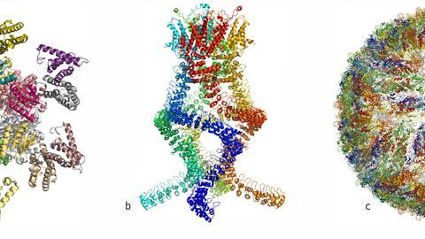
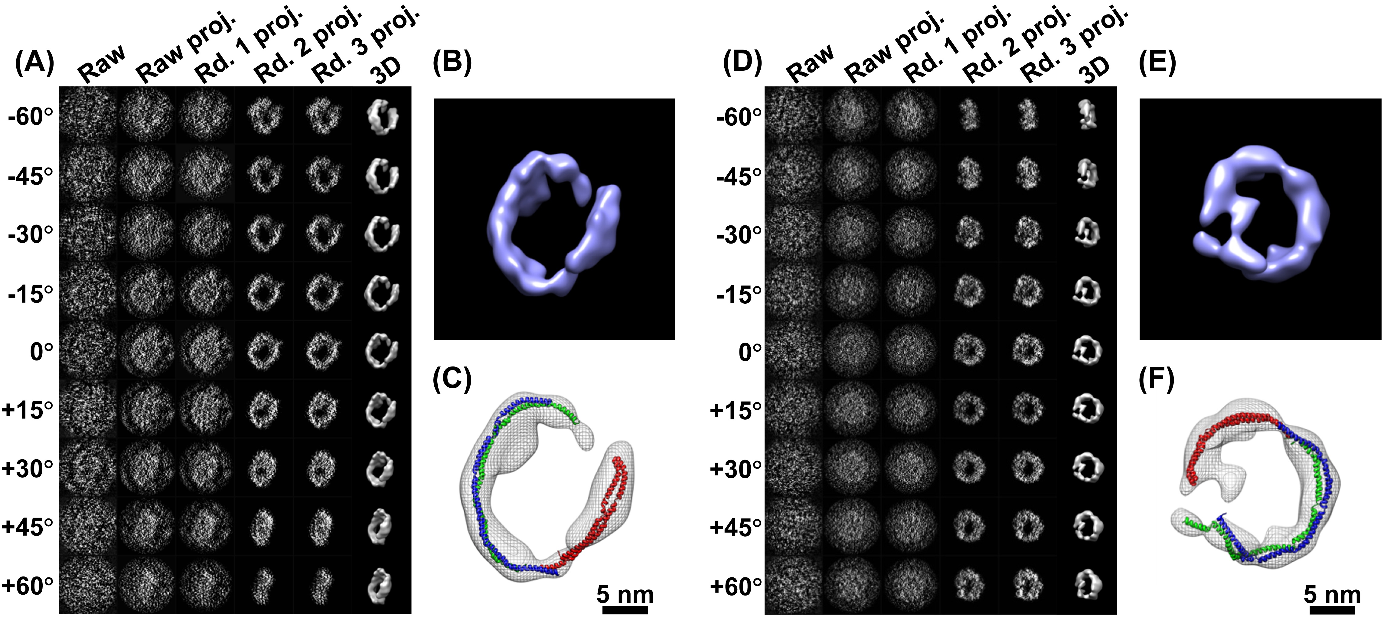
Comparison of reported structures from electron microscopy of TRP... |  Download Scientific Diagram
Comparison of reported structures from electron microscopy of TRP... | Download Scientific Diagram

Under the Electron Microscope – A 3-D Image of an Individual Protein -  Berkeley Lab – News Center
Under the Electron Microscope – A 3-D Image of an Individual Protein - Berkeley Lab – News Center

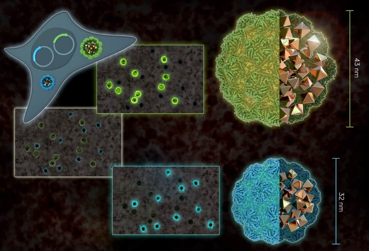
Bacterial nanostructures act as electron-microscope-compatible gene  reporters – Physics World
Bacterial nanostructures act as electron-microscope-compatible gene reporters – Physics World

Proteins under the microscope. A new device called the Volta phase… | by  eLife | Life's Building Blocks | Medium
Proteins under the microscope. A new device called the Volta phase… | by eLife | Life's Building Blocks | Medium